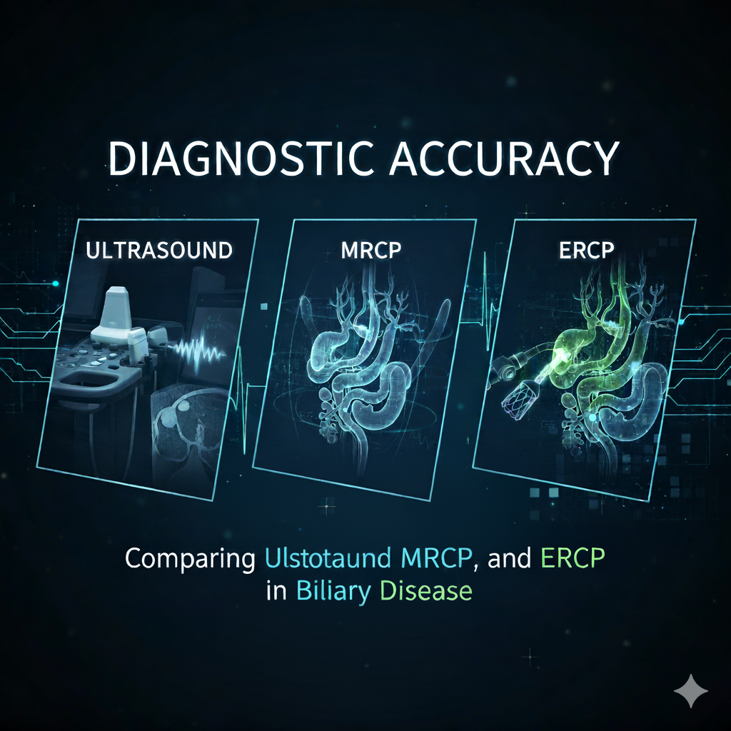
Diagnostic Accuracy: Comparing Ultrasound, MRCP, and ERCP in Biliary Disease
Accurate diagnosis of biliary tract disease remains fundamental to effective patient management and optimal therapeutic outcomes. As diagnostic imaging continues to evolve, clinicians must understand the comparative strengths, limitations, and appropriate applications of available modalities. This comprehensive analysis examines the diagnostic accuracy of transabdominal ultrasound (US), magnetic resonance cholangiopancreatography (MRCP), and endoscopic retrograde cholangiopancreatography (ERCP) in evaluating biliary pathology, providing evidence-based insights to guide clinical decision-making.
Understanding Biliary Disease: A Diagnostic Challenge
Biliary tract disorders encompass a spectrum of conditions ranging from choledocholithiasis (common bile duct stones) and benign strictures to malignant obstructions and congenital anomalies. Choledocholithiasis affects approximately 10% of patients with symptomatic gallstones and represents the most common cause of biliary obstruction. Accurate identification of common bile duct (CBD) stones is essential to avoid the substantial morbidity and mortality that can result from undetected biliary obstruction.
The clinical presentation of biliary disease varies widely, with symptoms including jaundice, right upper quadrant pain, fever, abnormal liver function tests, and in severe cases, acute cholangitis or pancreatitis. Given this heterogeneity, selecting the optimal diagnostic imaging pathway requires careful consideration of test performance characteristics, patient-specific factors, disease prevalence, therapeutic intent, and cost-effectiveness.
Transabdominal Ultrasound: The First-Line Imaging Modality
Diagnostic Performance
Transabdominal ultrasound serves as the initial imaging modality for suspected biliary disease due to its widespread availability, non-invasive nature, absence of ionizing radiation, and relatively low cost. However, its diagnostic accuracy for detecting CBD stones demonstrates significant limitations.
Multiple large-scale studies have consistently shown that ultrasound has limited sensitivity for CBD stone detection, ranging from 32% to 73%, though it maintains high specificity approaching 100%. In a landmark study by Varghese et al. involving 191 patients, ultrasound demonstrated a sensitivity of only 38%, specificity of 100%, and overall diagnostic accuracy of 89% for choledocholithiasis when compared to direct cholangiography.
A comprehensive review of Pakistani medical literature similarly confirmed ultrasound’s suboptimal performance, with sensitivity of 50%, specificity of 96.4%, and positive predictive value of 80.5% for detecting CBD stones. These findings underscore that normal ultrasound findings can be falsely reassuring, as demonstrated by studies showing that among 71 patients with CBD stones confirmed on MRCP, 30 had entirely normal ultrasound examinations.
Strengths and Limitations
Ultrasound excels in several important areas:
CBD Dilatation Detection: Ultrasound reliably detects a dilated CBD with sensitivity up to 87%, making it valuable for initial assessment of biliary obstruction
Real-Time Imaging: Dynamic evaluation allows assessment of gallbladder contractility and real-time visualization during provocative maneuvers
Point-of-Care Accessibility: Bedside ultrasound provides immediate diagnostic information in acute clinical settings
Safety Profile: No radiation exposure or contrast administration makes it ideal for pregnant patients and those with renal dysfunction
Cost-Effectiveness: As the least expensive imaging modality, ultrasound remains appropriate for initial screening
However, significant limitations restrict ultrasound’s diagnostic utility:
Poor Distal CBD Visualization: Bowel gas and overlying duodenum frequently obscure the distal common bile duct, precisely where stones commonly lodge
Small Stone Detection: Stones less than 5mm frequently escape ultrasound detection
Operator Dependence: Diagnostic accuracy varies substantially based on sonographer experience and skill level
Body Habitus Limitations: Obesity significantly degrades image quality and diagnostic confidence
Inability to Visualize Pancreatic Duct: Ultrasound provides no information about pancreatic ductal pathology
Magnetic Resonance Cholangiopancreatography: The Non-Invasive Gold Standard
Diagnostic Performance for Choledocholithiasis
MRCP has emerged as the premier non-invasive imaging modality for biliary and pancreatic ductal evaluation, offering exceptional diagnostic accuracy without the risks associated with invasive procedures. MRCP demonstrates sensitivity ranging from 80% to 96.2%, specificity from 91.8% to 100%, and overall diagnostic accuracy between 89% and 97% for detecting choledocholithiasis.
A Pakistani study conducted at Services Hospital Lahore involving 170 patients reported MRCP sensitivity of 83.33%, specificity of 93.88%, and diagnostic accuracy of 89.41% for choledocholithiasis detection, with per-operative findings serving as the gold standard. International studies have reported even higher accuracy, with Varghese et al. demonstrating MRCP sensitivity of 91%, specificity of 98%, and diagnostic accuracy of 97% compared to direct cholangiography.
A comprehensive analysis involving 258 patients randomized to either MRCP or ERCP demonstrated that MRCP achieved 96% sensitivity for detecting bile duct obstruction with 89% overall diagnostic accuracy—identical to ERCP’s performance. These findings establish MRCP as having diagnostic accuracy comparable to invasive cholangiography for most biliary pathologies.
Performance Across Various Biliary Conditions
Choledocholithiasis: MRCP excels in detecting CBD stones, particularly those exceeding 6mm in diameter. Studies excluding stones less than 6mm reported sensitivity, specificity, and accuracy of 100%, 99%, and 99%, respectively. However, MRCP may underestimate the total number of stones present, particularly small calculi (less than 4mm).
Biliary Strictures: For differentiating malignant from benign biliary strictures, MRCP demonstrates sensitivity of 81% and specificity of 70%, comparable to ERCP’s 74% sensitivity and 70% specificity. A lengthy segment of extrahepatic bile duct stricture with irregular margins and asymmetric narrowing suggests cholangiocarcinoma, whereas a short segment with regular margins and symmetric narrowing suggests benign etiology, regardless of imaging modality.
Malignant Obstruction: In diagnosing pancreaticobiliary malignancies, MRCP shows sensitivity ranging from 81% to 94% and specificity from 92% to 100%, with positive likelihood ratios between 10.12 and 43. MRCP provides comprehensive visualization of mass lesions, vascular involvement, and metastatic disease that inform surgical planning and resectability assessment.
Pancreatic Duct Pathology: Unlike ultrasound and even ERCP (which may fail due to difficult cannulation), MRCP consistently visualizes the entire pancreatic ductal system, detecting strictures, dilatation, intraductal stones, and ductal disruption.
Advantages of MRCP
MRCP offers numerous advantages that have established it as the preferred diagnostic modality for suspected biliary disease:
Non-Invasive: MRCP requires no endoscopic intervention, eliminating risks of pancreatitis, perforation, bleeding, and sedation-related complications.
No Ionizing Radiation: Unlike CT and ERCP, MRCP uses magnetic fields and radio waves, making it safe for younger patients and those requiring repeated imaging.
No Contrast Administration: Standard MRCP sequences do not require intravenous contrast, avoiding risks of contrast-induced nephropathy and allergic reactions.
Comprehensive Visualization: MRCP provides complete visualization of intrahepatic and extrahepatic bile ducts, pancreatic duct, gallbladder, and surrounding structures in a single examination.
Independent of Anatomy: Unlike ERCP, which may fail due to prior gastric surgery, duodenal diverticula, or ampullary tumors, MRCP successfully images the biliary tree regardless of anatomical variations.
Failed ERCP Alternative: MRCP serves as an excellent option for patients with unsuccessful or contraindicated ERCP procedures.
Patient Preference: Studies demonstrate that patients overwhelmingly prefer MRCP to diagnostic ERCP when given the choice.
Limitations of MRCP
Despite its exceptional performance, MRCP has several important limitations:
Small Stone Detection: MRCP sensitivity decreases for stones less than 5mm in diameter, potentially missing clinically significant small calculi.
Stone Number Underestimation: MRCP tends to detect fewer stones than ERCP and may not accurately represent the total stone burden, particularly when multiple small stones are present.
Contraindications: Patients with cardiac pacemakers, certain metallic implants, cochlear implants, and severe claustrophobia cannot undergo MRI examinations.
Limited Availability: In many regions, particularly in Pakistan, MRCP availability remains restricted to tertiary care centers, limiting access for rural populations.
Cost Considerations: MRCP costs approximately £650 (or equivalent) per examination, substantially higher than ultrasound though lower than therapeutic ERCP with complications.
Motion Artifacts: Patient movement or inability to hold breath during image acquisition degrades image quality and diagnostic confidence.
Limited Tissue Diagnosis: Unlike ERCP, MRCP cannot obtain tissue specimens for histopathologic confirmation of suspected malignancies.
Endoscopic Retrograde Cholangiopancreatography: Therapeutic Capability with Diagnostic Risks
Diagnostic Performance
ERCP has historically served as the gold standard for biliary tract evaluation, offering direct visualization through contrast opacification of the biliary and pancreatic ductal systems. ERCP demonstrates sensitivity exceeding 90% and specificity approaching 100% for most biliary pathologies.
In comparative studies, ERCP showed sensitivity of 90% for choledocholithiasis detection, with overall diagnostic accuracy of 97.5% for malignant biliary obstruction. A prospective randomized trial involving 258 patients demonstrated ERCP’s 89% overall accuracy for correct diagnosis—identical to MRCP’s performance.
However, ERCP’s diagnostic accuracy depends critically on successful cannulation of the ampulla of Vater and adequate opacification of the biliary tree. Cannulation failure occurs in 3-10% of cases, particularly in patients with periampullary diverticula, previous gastric surgery, or ampullary tumors. In studies where cannulation failed, ERCP provided no diagnostic information while still exposing patients to procedural risks.
The Shift from Diagnostic to Therapeutic ERCP
The advent of high-quality non-invasive imaging modalities, particularly MRCP and endoscopic ultrasound (EUS), has fundamentally transformed ERCP’s role in clinical practice. ERCP is no longer recommended as a purely diagnostic procedure but should be reserved for cases requiring therapeutic intervention.
Current evidence-based guidelines emphasize that indiscriminate use of diagnostic ERCP results in an increasing proportion of patients in whom therapeutic intervention proves unnecessary, exposing them to significant procedural risks without benefit. Modern practice dictates that ERCP should be performed only when there is high clinical suspicion for biliary disease requiring endoscopic treatment, such as:
Stone extraction via basket retrieval or lithotripsy
Biliary sphincterotomy for stone clearance or sphincter of Oddi dysfunction
Biliary stent placement for malignant or benign strictures
Tissue sampling via brush cytology or biopsy forceps
Drainage of bile duct abscesses or fluid collections
Management of bile leaks following surgery or trauma
Complications and Mortality
ERCP carries substantial risks that must be carefully weighed against potential benefits. Overall complication rates range from 5% to 10%, with mortality rates between 0.2% and 1.0%.
Post-ERCP Acute Pancreatitis represents the most common complication, occurring in approximately 2.3% to 10% of procedures. Most cases are mild and resolve with conservative management, but severe pancreatitis develops in 1-3% of patients, with mortality rates reaching 20-30% when multiple organ dysfunction occurs. Risk factors for post-ERCP pancreatitis include:
Difficult or failed cannulation attempts
Sphincter of Oddi dysfunction
Female gender
Young age
Absence of chronic pancreatitis
Repeated contrast injection into the pancreatic duct
Pancreatic sphincterotomy
Perforation occurs in approximately 1% of ERCP procedures, with mortality rates of 16-18% when perforation is recognized. Bleeding from sphincterotomy or biopsy sites carries a 30-40% mortality rate when major vessel injury occurs.
Infectious Complications, including acute cholangitis and cholecystitis, result from inadequate biliary drainage or introduction of bacteria during the procedure. Cardiovascular Complications, though rare (1% incidence), can be life-threatening in patients with significant comorbidities.
Recent population-based data from patients with chronic liver disease revealed in-hospital mortality of 3.03% following ERCP compared to 1.58% in patients without liver disease, demonstrating the profound impact of underlying comorbidities on ERCP outcomes.
Time to death following ERCP-related complications is alarmingly short, with median time from ERCP to death of only 9.5 days in fatality cases compared to 40 days in control patients. This underscores the importance of rigorous patient selection and the need for alternative diagnostic approaches in appropriate clinical scenarios.
Endoscopic Ultrasound: The Emerging Diagnostic Alternative
Endoscopic ultrasound (EUS) has emerged as an excellent minimally invasive alternative to ERCP for diagnosing biliary obstruction, combining endoscopic visualization with high-frequency ultrasound imaging. EUS demonstrates diagnostic accuracy of 89.5% to 94.3% for choledocholithiasis, with sensitivity of 89.5% to 100% and specificity of 96.5% to 96.6%.
EUS proves particularly valuable for detecting small stones and bile sludge that may escape detection by other modalities. The sensitivity and specificity of EUS for detecting CBD stones exceeding 4mm reach 96% and 100%, respectively. EUS can reliably differentiate patients who require therapeutic ERCP from those who do not, potentially avoiding 15-30% of unnecessary ERCP procedures.
Studies comparing EUS directly with MRCP in the same patients have shown mixed results, with some demonstrating EUS superiority (particularly for small stones) while others show comparable performance. EUS accuracy is highly operator-dependent, requiring substantial training and experience to achieve optimal results.
Clinical Decision-Making: Selecting the Appropriate Diagnostic Pathway
Risk Stratification and Patient Selection
The American Society for Gastrointestinal Endoscopy (ASGE) guidelines recommend risk-stratifying patients into low, intermediate, and high probability categories for choledocholithiasis based on clinical, laboratory, and ultrasound findings.
High-Risk Criteria (probability >50%) include:
CBD stone visualized on ultrasound
Clinical ascending cholangitis
Total bilirubin >4 mg/dL with dilated CBD on imaging
Intermediate-Risk Criteria (probability 10-50%) include:
Abnormal liver biochemistry other than isolated bilirubin elevation
Age >55 years
Dilated CBD on ultrasound (>6mm with gallbladder in situ)
Low-Risk Criteria (probability <10%):
Normal laboratory values
Non-dilated CBD on imaging
No other risk factors
Management recommendations based on risk stratification:
Low-Risk Patients: Proceed directly to laparoscopic cholecystectomy without preoperative biliary imaging
High-Risk Patients: Proceed directly to ERCP for combined diagnosis and therapy
Intermediate-Risk Patients: Perform MRCP or EUS for further evaluation; reserve ERCP for those with positive findings requiring intervention
Cost-Effectiveness Analysis
Multiple cost-effectiveness analyses have demonstrated that MRCP represents a dominant strategy (both more effective and less costly) compared to ERCP in patients with low to moderate probability of choledocholithiasis.
At disease prevalence below 60%, MRCP proves less costly than initial ERCP; above this threshold, ERCP becomes the more economical approach. Probabilistic sensitivity analysis indicates an 83% likelihood that MRCP has a cost-effectiveness ratio more favorable than $50,000 per quality-adjusted life year (QALY) gained.
In Pakistan, where MRCP costs substantially less than in Western healthcare systems and ERCP complications may incur catastrophic expenses for families, MRCP-first strategies offer even greater cost-effectiveness advantages. A recent Pakistani study demonstrated that contrast-enhanced CT provides diagnostic accuracy of 96.2% for biliary pathologies at lower cost than MRCP, suggesting it may represent another viable alternative in resource-constrained settings.
Proposed Diagnostic Algorithm
Based on current evidence and international guidelines, we recommend the following diagnostic algorithm for suspected biliary disease:
Step 1: Initial Evaluation
Comprehensive history and physical examination
Liver function tests (bilirubin, alkaline phosphatase, ALT, AST)
Complete blood count
Transabdominal ultrasound
Step 2: Risk Stratification
Apply ASGE criteria to categorize patients as low, intermediate, or high risk
Step 3: Pathway Selection
For Low-Risk Patients:
Proceed to cholecystectomy without additional imaging
Consider intraoperative cholangiography if available
For High-Risk Patients:
Proceed directly to ERCP for combined diagnosis and therapy
Ensure therapeutic intent and preparedness for intervention
For Intermediate-Risk Patients:
Obtain MRCP as first-line advanced imaging (if available)
Consider EUS as alternative if MRCP contraindicated or unavailable
Reserve ERCP for patients with positive MRCP/EUS requiring therapeutic intervention
Step 4: Follow-Up
Persistently elevated liver function tests despite normal imaging warrant repeat evaluation
Clinical symptoms inconsistent with imaging findings require additional investigation
Special Considerations for the Pakistani Healthcare Context
Healthcare delivery in Pakistan presents unique challenges that influence diagnostic pathway selection. Limited MRCP availability in many regions, financial constraints for patients, and variable access to advanced endoscopic services require practical adaptations to international guidelines.
Ultrasound remains the cornerstone of initial biliary evaluation throughout Pakistan due to universal availability and minimal cost. However, clinicians must recognize its limitations and maintain a low threshold for advanced imaging when clinical suspicion persists despite normal ultrasound findings.
MRCP availability has expanded in major cities including Lahore, Karachi, Islamabad, and Rawalpindi, though access remains limited in rural areas. The development of indigenous medical technology, such as Pakistan’s first locally manufactured FDR X-ray machine, demonstrates the country’s growing capacity for advanced medical equipment production that may extend to MRI systems in the future.
ERCP expertise is concentrated in tertiary care centers, with pioneers such as the team at Aga Khan University Hospital in Karachi establishing teleradiology and training programs to expand access. However, complication management capabilities vary significantly across facilities, necessitating careful patient selection and appropriate referral patterns.
Contrast-enhanced CT represents an underutilized alternative that provides diagnostic accuracy comparable to MRCP (96.2%) while offering additional information about extra-biliary pathology at lower cost. CT should be considered when MRCP is unavailable, contraindicated, or financially prohibitive.
Conclusion: Evidence-Based Diagnostic Imaging Selection
The landscape of biliary imaging has evolved dramatically, with multiple modalities offering complementary strengths and limitations. Transabdominal ultrasound serves as an appropriate first-line screening tool despite limited sensitivity for CBD stones. MRCP has emerged as the preferred diagnostic modality for intermediate-risk patients, offering exceptional accuracy without invasive risks. ERCP should be reserved exclusively for patients requiring therapeutic intervention, given its substantial complication rates and mortality risk.
The key to optimal patient care lies not in selecting a single “best” test but in thoughtfully matching imaging modality to clinical context, disease probability, therapeutic intent, local expertise, and resource availability. Risk stratification using validated criteria allows clinicians to avoid unnecessary invasive procedures while ensuring timely intervention for patients who will benefit.
As Pakistan’s healthcare infrastructure continues to develop, expanding access to MRCP and EUS will allow more patients to benefit from accurate non-invasive diagnosis. Until then, judicious use of available resources, careful clinical assessment, and appropriate patient selection remain essential to delivering high-quality, cost-effective biliary disease management.
Understanding the comparative diagnostic accuracy of ultrasound, MRCP, and ERCP empowers clinicians to make evidence-based decisions that optimize patient outcomes while minimizing risks and costs—the fundamental goals of modern diagnostic imaging.
Cum sociis natoque penatibus et magnis dis parturient montes nascetur ridiculus mus. Donec quam felis ultricies nec pellentesque eu pretium quis sem.