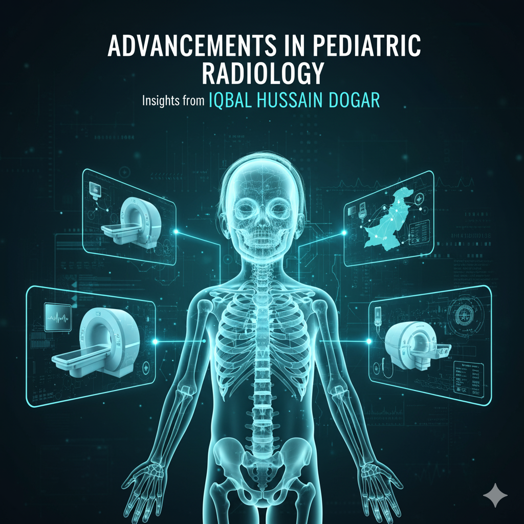
Advancements in Pediatric Radiology: Insights from Dr. Iqbal Hussain Dogar
Pediatric radiology has undergone remarkable transformations in recent years, driven by technological innovations, enhanced safety protocols, and dedicated leadership from visionary professionals. Dr. Iqbal Hussain Dogar, Principal of Gujranwala Medical College and Allied Teaching Hospitals and a distinguished Professor of Pediatric Radiology, stands at the forefront of these advancements, bringing international recognition to Pakistan’s contributions in this specialized field.
The Evolution of Pediatric Radiology: A Global Perspective
The global pediatric radiology market has experienced substantial growth, valued at USD 6.39 billion in 2023 and projected to reach USD 9.93 billion by 2032, with a compound annual growth rate of 5.02%. This expansion reflects the increasing demand for specialized diagnostic imaging services tailored to children’s unique physiological needs and the integration of cutting-edge technologies that prioritize both diagnostic accuracy and patient safety.
Recent developments in imaging techniques have revolutionized how clinicians approach pediatric diagnosis and treatment. Advanced modalities including low-dose radiation imaging equipment, artificial intelligence-enhanced image analysis, and minimally invasive diagnostic tools have collectively improved children’s safety while ensuring more efficient and accurate diagnoses.
Dr. Iqbal Hussain Dogar: A Pioneer in Pediatric Radiology
With an impressive academic foundation comprising MBBS, DMRD (Radiology), FCPS (Radiology), and M.Phil in Public Health, Dr. Dogar has dedicated his career to advancing medical education and pediatric healthcare delivery. His professional journey spans over two decades, including significant tenures as Associate Professor and Professor of Pediatric Radiology at prestigious institutions such as King Edward Medical University, The Children’s Hospital Lahore, and Faisalabad Medical University.
Groundbreaking Contributions: The Vascular Anomaly Centre
One of Dr. Dogar’s most transformative achievements has been the establishment of the Vascular Anomaly Centre at Gujranwala Medical College. This pioneering initiative addresses the critical need for specialized treatment of vascular anomalies in Pakistan, a country with limited healthcare resources. In February 2025, Dr. Dogar presented this transformative project at the European Congress of Radiology (ECR) in Vienna, Austria, where it was recognized as a “Beacon of Hope for Transforming Lives”.
The Vascular Anomaly Centre represents a paradigm shift in how complex vascular conditions are diagnosed and managed in pediatric patients. Dr. Dogar’s work in this area, including his presentation on “Pediatric Neurovascular Intracranial Vascular Anomalies and Their Endovascular Management” at the IRSP Annual Meeting in Karachi, demonstrates his commitment to minimally invasive procedures and advanced interventional radiology techniques.
Radiation Dose Reduction: Protecting Young Patients
One of the most critical advancements in pediatric radiology involves minimizing radiation exposure while maintaining diagnostic quality. Children are two to three times more susceptible to potential harmful effects of ionizing radiation than adults, making dose optimization particularly crucial.
Low-Dose CT Imaging Techniques
Modern pediatric CT protocols employ several innovative approaches to reduce radiation exposure. Reducing tube voltage from 120 kVp to 100 kVp or 80 kVp can theoretically yield dose reductions of 33% and 65%, respectively. Advanced reconstruction techniques, including:
Adaptive Statistical Iterative Reconstruction (ASIR): Reduces CT dose index by 8% and dose length product by 30%
Model-Based Iterative Reconstruction (MBIR): Offers enhanced noise reduction and low-contrast detectability
Deep Learning-Based Reconstruction: Enables radiation dose reductions of 36-70% while maintaining diagnostic accuracy
The Image Gently Alliance, a coalition of healthcare organizations dedicated to pediatric imaging safety, has established comprehensive guidelines and protocols to ensure radiation doses are optimized at the point of care. Their initiative emphasizes the ALARA (As Low As Reasonably Achievable) principle, which has become the gold standard in pediatric imaging practices worldwide.
Photon-Counting Detector CT: The Next Generation
Photon-counting detector computed tomography (PCD-CT) represents the most significant technological advancement in pediatric imaging. Unlike traditional energy-integrating detectors that convert X-rays into visible light, PCD-CT directly transforms X-ray photons into electrical signals, offering multiple advantages:
Superior spatial resolution through smaller detector elements (0.151 mm × 0.176 mm versus 0.45 mm × 0.51 mm in conventional CT)
Elimination of electronic noise, improving image quality at lower radiation doses
Enhanced scan speed up to 74 cm/s, reducing the need for sedation in pediatric patients
Radiation dose reduction of 31-47% while maintaining or improving image quality
St. Louis Children’s Hospital reported that in 2023, with the implementation of photon-counting CT technology, 96.5% of pediatric patients could be scanned without any sedation—a remarkable achievement that significantly improves patient safety and comfort.
Artificial Intelligence: Transforming Pediatric Radiology
Artificial intelligence has emerged as a critical decision support tool for pediatric radiologists, offering valuable assistance in classifying lesions, predicting prognosis, and treatment response planning. AI applications in pediatric neuroradiology are particularly promising, addressing unique challenges such as continuously developing brains with age-specific anatomical and physiological changes.
AI-Driven Dose Optimization
Deep convolutional neural networks (CNNs) have become the most common AI technique for dose optimization in pediatric radiology, enabling 36-70% radiation dose reduction without losing diagnostic information. AI algorithms can analyze ultra-low-dose CT and low-quality MRI exams to produce high-quality diagnostic images, making previously challenging scans more accessible and safer for pediatric patients.
Accelerating Image Acquisition
AI reduces MRI and CT scanner time by employing advanced deep learning algorithms to accelerate image acquisition through compressed sensing and undersampling, producing diagnostic-quality images with significantly fewer data points and in shorter timeframes. This is particularly beneficial for pediatric patients who struggle to remain still during lengthy examinations.
Whole-Body MRI: Radiation-Free Comprehensive Imaging
Whole-body MRI (WBMRI) has expanded rapidly over the past decade as a radiation-free imaging method for evaluating multi-system diseases in children. Current indications include:
Screening for presymptomatic lesions in cancer predisposition syndromes
Tumor staging in known malignancies
Investigating fevers of unknown origin
Diagnosing and monitoring rheumatologic diseases and vascular anomalies
WBMRI protocols typically employ coronal and sagittal short tau inversion recovery (STIR) imaging combined with diffusion-weighted imaging (DWI), which has proven particularly valuable for pediatric oncology applications. The technique has demonstrated diagnostic performance comparable to PET/CT for initial staging of pediatric lymphoma, offering a radiation-free alternative.
Ultrasound Innovations: Safe and Accessible Imaging
Ultrasound technology continues to evolve with pediatric-specific innovations that expand diagnostic capabilities while maintaining its inherent safety advantages.
Contrast-Enhanced Ultrasound (CEUS)
Contrast-enhanced ultrasound has become a game-changer in pediatric imaging, particularly for focal liver lesion evaluation, blunt abdominal trauma assessment, and various organ-specific applications. The FDA approved ultrasound contrast agents for intravenous use in children for characterizing focal liver lesions and during echocardiography, with off-label applications rapidly expanding.
CEUS offers numerous advantages over CT and MRI:
No ionizing radiation exposure
No need for sedation or general anesthesia
Excellent safety profile with no risk of kidney or liver damage
Real-time evaluation of blood flow and tissue perfusion
Portability for bedside examinations
Cost-effectiveness compared to CT and MRI
Advanced Ultrasound Techniques
Recent innovations include microvascular flow imaging (MFI) for enhanced visualization of tumor vascularity, ultrasound-derived fat fraction (UDFF) for quantitative hepatic fat assessment, and elastography for tissue characterization. These technologies mirror adult applications while offering unique benefits when applied to pediatric patients.
MRI Safety: Reducing the Need for Sedation
Young children often require sedation or general anesthesia during MRI examinations to minimize motion artifacts. However, there are increasing concerns regarding the effects of sedation on developing brains, driving innovation in sedation-free imaging strategies.
Non-Pharmacological Interventions
Meta-analysis of 20 studies involving 33,873 patients demonstrated that nonpharmacological interventions reduce the need for sedation and general anesthesia in children aged 2-18 years undergoing MRI (risk ratio: 0.68 for randomized controlled trials and 0.58 for non-randomized studies). Effective strategies include:
Mock MRI training and preparation programs
Video goggles and distraction techniques
Feed-and-swaddle technique for infants
Child life specialist involvement
Parental presence during examinations
These interventions are most effective in children aged 3-10 years, achieving success rates as high as 86% in avoiding sedation while maintaining diagnostic image quality.
Ultra-Fast MRI Sequences
Technical advances including T2-weighted turbo-spin-echo PROPELLER for brain imaging and 4D free-breathing MRI techniques for abdominal imaging have significantly reduced scan times while improving image quality. These motion-robust sequences minimize the need for sedation while ensuring accurate diagnosis.
Dr. Dogar’s Educational Leadership and Research Impact
Beyond his clinical contributions, Dr. Dogar has demonstrated exceptional leadership in medical education. Under his principalship, Gujranwala Medical College has implemented numerous transformative initiatives, including:
Establishment of a Surgical ICU and Pain Management Clinic
Inauguration of the Inclusive Health Society
Creation of a Student’s Centre for empowerment
Addition of a 502-bedded teaching hospital with advanced equipment
His commitment to implementing integrated modular curriculum and advancing healthcare infrastructure reflects a comprehensive vision for elevating medical education standards in Pakistan. Dr. Dogar’s research publications span various aspects of diagnostic radiology, including comparative studies of ultrasonographic findings, magnetic resonance cholangiopancreatography, and depressed skull fracture evaluation.
Future Directions in Pediatric Radiology
The future of pediatric radiology promises continued innovation across multiple fronts:
Federated Learning and Collaborative AI Development
Federated learning allows AI models to learn from diverse data across multiple institutions without compromising patient privacy, addressing the critical challenge of pediatric data scarcity. This collaborative approach enhances AI model robustness and generalizability, particularly important given that children represent less than 1% of available data in public medical imaging datasets.
Hybrid Imaging Technologies
PET-MRI fusion systems have overcome technical barriers, achieving simultaneous acquisition with improved silicon photomultiplier detectors. This hybrid modality quantifies tumor metabolism while mapping anatomical boundaries, reducing prostate cancer biopsy false negatives by 28% compared to multiparametric MRI alone.
4D Imaging and Real-Time Visualization
The transition from 3D to 4D imaging enables real-time visualization of moving structures, with temporal resolution improvements revolutionizing cardiac and vascular assessments. Four-dimensional CT perfusion imaging has become standard in acute stroke management, generating whole-brain perfusion maps in 2.3 seconds.
Conclusion: A Vision for the Future
The advancements in pediatric radiology exemplified by Dr. Iqbal Hussain Dogar’s pioneering work represent a convergence of technological innovation, clinical expertise, and unwavering commitment to patient safety. His establishment of the Vascular Anomaly Centre, presentations at international conferences, and leadership in medical education have positioned Pakistan as a contributor to global pediatric radiology excellence.
As imaging technologies continue to evolve with photon-counting CT, artificial intelligence integration, advanced ultrasound techniques, and radiation-free whole-body MRI, the future of pediatric radiology promises earlier disease detection, more precise diagnoses, and improved outcomes for young patients worldwide. Dr. Dogar’s transformative leadership and dedication to advancing pediatric healthcare serve as inspiration for the next generation of radiologists committed to imaging children gently, wisely, and with the highest standards of care.
The field of pediatric radiology stands at an exciting juncture where cutting-edge technology meets compassionate care, ensuring that every child receives safe, accurate, and effective diagnostic imaging tailored to their unique developmental needs.
Donec vitae sapien ut libero venenatis faucibus. Nullam quis ante. Etiam sit amet orci eget eros faucibus tincidunt. Duis leo. Sed fringilla mauris sit amet nibh. Donec sodales sagittis magna. Sed consequat, leo eget bibendum sodales, augue velit cursus nunc, quis gravida magna mi a libero.